retrolisthesis.webp

Retrolisthesis

Questions

WHAT IS RETROLISTHESIS?

Retrolisthesis is a spinal condition where one vertebra slips backward relative to the vertebra below it. This backward displacement can lead to spinal instability, nerve compression, and a variety of symptoms depending on the location and severity of the slippage.

WHAT ARE THE SYMPTOMS OF RETROLISTHESIS?

The symptoms of retrolisthesis can vary based on the affected region of the spine (cervical, thoracic, or lumbar) and the extent of the displacement. Common symptoms include:

1: Pain: Localized pain at the site of the slippage, which can radiate to the limbs if nerve roots are compressed.
2: Numbness and Tingling: Often in the arms or legs, depending on the location of the retrolisthesis.
3: Muscle Weakness: Weakness in the affected limbs due to nerve compression.
4: Reduced Mobility: Stiffness and decreased range of motion in the spine.
5: Postural Changes: Visible changes in posture, such as a noticeable curve in the spine or an altered gait.
6: Chronic Pain: Persistent pain in the neck or back, which can be aggravated by activity.
7: Headaches: Especially if the cervical spine is affected.
8: Functional Impairment: Difficulty performing daily activities due to pain and reduced mobility.

WHAT ARE THE CAUSES FOR RETROLISTHESIS?

Several factors can contribute to the development of retrolisthesis, including:

1: Degenerative Changes: Age-related degeneration of the spine, such as disc degeneration and facet joint arthritis.
2: Trauma: Acute injuries or fractures to the spine.
3: Congenital Defects: Structural abnormalities present from birth.
4: Post-Surgical Changes: Complications or changes following spinal surgery.
5: Chronic Stress or Overuse: Repetitive movements or heavy lifting that strain the spine.
6: Spinal Conditions: Conditions like osteoporosis, spondylosis, or spinal tumors that weaken the spine's structural integrity.

Pathology: The pathology of retrolisthesis involves several anatomical and physiological changes:
1: Vertebral Slippage: A backward displacement of one vertebra relative to the vertebra below it. The degree of slippage can be classified as mild, moderate, or severe.
2: Disc Degeneration: The intervertebral discs may become worn or degenerate, reducing their ability to cushion the vertebrae effectively. This degeneration can contribute to instability and slippage.
3: Facet Joint Dysfunction: The facet joints, which help stabilize the vertebrae, may become arthritic or misaligned, contributing to spinal instability and backward displacement.
4: Ligamentous Instability: The ligaments supporting the spine may become lax or weakened, contributing to the slippage and instability of the vertebrae.
5: Spinal Canal Narrowing: Retrolisthesis can reduce the diameter of the spinal canal, leading to spinal stenosis, which compresses the spinal cord or nerve roots and results in pain, numbness, and other neurological symptoms.
6: Nerve Impingement: The backward displacement can compress or pinch the nerves exiting the spinal column, causing pain, numbness, tingling, and muscle weakness in the affected areas.

DIAGNOSIS OF RETROLISTHESIS.

Physical Examination: Evaluation of lower back pain, stiffness, muscle weakness, and neurological deficits.

Range of Motion Testing: Examination of the spine's flexibility and any limitations in movement.

Neurological Examination: Assessment of reflexes, muscle strength, sensation, and coordination to identify signs of nerve compression or damage.

Standard X-rays: Provide detailed images of the spine's bone structures, allowing visualization of vertebral alignment and degree of slippage.

Lateral Views: Side-view X-rays are particularly useful for assessing the alignment and degree of displacement of the vertebrae.

Flexion and Extension X-rays: Taken while the patient bends forward and backward to evaluate the stability and dynamic movement of the spine.

Magnetic Resonance Imaging (MRI): MRI provides detailed images of soft tissues, including intervertebral discs, spinal cord, and nerve roots. Helps identify any nerve compression or spinal cord involvement, which is critical for planning treatment.

Computed Tomography (CT) Scan: CT scan offers detailed cross-sectional images of the spine, useful for assessing bone structures and the degree of slippage. It can create 3D images of the spine for a comprehensive assessment.

Electromyography (EMG) and Nerve Conduction Studies (NCS): EMG and NCS measure the electrical activity of muscles and the speed of nerve signals, helping to identify nerve damage or compression related to retrolisthesis.

Bone Scans: A bone scan is used if there is suspicion of an underlying condition such as a fracture, infection, or tumor. It helps identify areas of high bone metabolism.

Discography: Involves injecting a contrast dye into the intervertebral disc to assess its integrity and pain response. Helps identify problematic discs that may contribute to retrolisthesis.

TREATMENT FOR RETROLISTHESIS.

Medication: Non-steroidal anti-inflammatory Drugs (NSAIDs), analgesics, muscle relaxants, opioids, corticosteroids, anticonvulsants and anti-depressants, etc.
(Note: Medication should not be taken without the doctor’s prescription.)

Surgeries:
When conservative treatments fail, or if there is significant nerve compression or spinal instability, surgical intervention may be necessary. Common surgical options include:

Spinal Fusion:
Spinal fusion is a process of using two or more vertebrae together to stabilize the spine.
   Types:
   a) Posterior Lumbar Interbody Fusion (PLIF): Access through the back.
   b) Anterior Lumbar Interbody Fusion (ALIF): Access through the front.
   c) Transforaminal Lumbar Interbody Fusion (TLIF): Posterior approach but more lateral than PLIF.

Laminectomy:
Laminectomy is the process of removal of part of the vertebral bone called the lamina.

Discectomy:
Discectomy is the process of removal of part or all of a herniated disc.

Foraminotomy:
Foraminotomy is the process of enlargement of the foramina (the openings where nerve roots exit the spine).

Artificial Disc Replacement:
Artificial disc replacement is the process of replacement of a damaged intervertebral disc with an artificial one.

WHAT IS THE PHYSIOTHERAPY TREATMENT RETROLISTHESIS?

Physiotherapists often use various electrical modalities to manage pain, reduce inflammation, and promote healing in patients with retrolisthesis:

Thermotherapy / Cryotherapy:
The use of heat/cold packs can help relieve pain and increase the range of motion..

Transcutaneous Electrical Nerve Stimulation (TENS):
TENS delivers low-voltage electrical currents through the skin to stimulate nerve fibers, which can help block pain signals to the brain and promote the release of endorphins. Electrodes are placed on the skin over the painful area, and the patient feels a tingling sensation.

Interferential Current Therapy (IFC):
IFT uses two high-frequency electrical currents that intersect deep within the tissues. The interference of these currents produces a low-frequency current that penetrates deeper into the tissues compared to TENS.
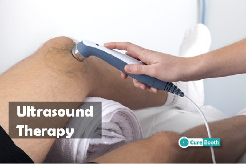
Ultrasound Therapy:
US helps promote tissue healing, reduce pain and inflammation. It uses high-frequency sound waves to produce deep heat within tissues, which increases blood flow, relaxes muscles, and promotes healing. The gel is applied to the skin, and a handheld ultrasound device is moved over the affected area.

Electromyographic Biofeedback (EMG):
EMG uses electrodes to detect muscle activity and provides feedback to the patient. This helps patients learn how to control muscle tension and improve muscle function. Electrodes are placed on the skin over the muscles, and the patient receives visual or auditory feedback.

Electrical Muscle Stimulation (EMS):
EMS helps in muscle strengthening and pain relief. It uses electrical currents to cause muscle contractions, which can help strengthen weak muscles, reduce muscle spasms, and improve blood circulation. Electrodes are placed on the skin over the target muscles, and the patient experiences muscle contractions.

Iontophoresis:
Iontophoresis deliver medication through the skin to reduce inflammation and pain. It uses a low electrical current to drive anti-inflammatory medications (e.g., corticosteroids) into the affected tissues. A medication-soaked pad is placed on the skin, and an electrode is attached to it to deliver the electrical current.

Microcurrent Therapy:
Microcurrent therapy is used for pain relief and tissue healing. It uses very low-level electrical currents to mimic the body's natural electrical currents, promoting cellular repair and reducing inflammation. Electrodes are placed on the skin, and the patient may feel a very mild or no sensation.

High-Voltage Pulsed Current (HVPC):
HVPC is used for pain relief, reduction of edema, and wound healing. It uses high-voltage electrical pulses to stimulate tissues, reduce swelling, and promote healing. Electrodes are placed on the skin over the affected area, and the patient feels a pulsing sensation.
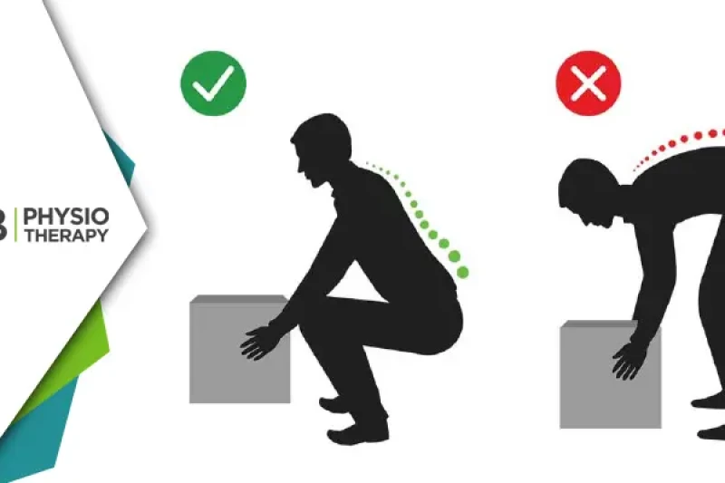
Exercise Therapy:
1: Strengthening Exercises: Focus on strengthening the core muscles, including the abdominal, back, and pelvic muscles, to provide better support for the spine, e.g. planks, bridges, pelvic tilts, and leg raises.
2: Flexibility Exercises: Stretching exercises to improve flexibility and reduce muscle tightness, e.g. hamstring stretches, hip flexor stretches, and lumbar stretches.
3: Stabilization Exercises: Exercises aimed at enhancing spinal stability and reducing the risk of further slippage, e.g., bird-dog, dead bug, and side planks.
4: Low-impact aerobic Exercises: Activities such as walking, swimming, or cycling to improve cardiovascular health without placing excessive stress on the spine.
5: Progressive Loading: This includes a gradual increase in intensity and slowly increasing the intensity and complexity of exercises as the patient progresses to build strength and endurance.

Manual Therapy:
Gentle movements are given to improve joint function and relieve pain. Soft tissue techniques like massage and myofascial release reduce muscle tension and improve blood flow.

Balance and Coordination Training:
Balance exercises help to improve stability and prevent falls, e.g. single-leg stands, balance board exercises, and stability ball exercises.

PATIENT EDUCATION.

Patient education is a vital component of managing retrolisthesis. Educating patients helps them understand their condition, adhere to treatment plans, and adopt lifestyle changes to prevent further injury and manage symptoms effectively. The patient is recommended ergonomic adjustments for workstations, including appropriate chair height, monitor level, and keyboard placement. Taught proper lifting techniques to avoid further injury, such as bending at the knees and keeping objects close to the body.

Retrolisthesis Physiotherapy Nearby

Select your City to find & connect with our experts regarding Physiotherapy for Retrolisthesis