gastrocnemius-rupture.webp

Gastrocnemius Rupture

Questions

WHAT IS GASTROCNEMIUS RUPTURE?

A Gastrocnemius rupture /tear is an injury to the gastrocnemius muscle, which is the larger and more superficial of the two muscles that make up the calf (the other being the soleus). This muscle is responsible for plantarflexion of the foot (pointing the toes downward) and plays a significant role in movements like running, jumping, and pushing off the ground.

Grades of Injury:
Grade I: Mild strain with minimal damage to the muscle fibers.
Grade II: Partial tear with more significant damage and greater loss of function.
Grade III: Complete rupture of the muscle, often requiring surgical intervention.

WHAT ARE THE CAUSES OF GASTROCNEMIUS RUPTURE?

A Gastrocnemius rupture/ tear is usually caused by sudden or forceful movements that overstretch the muscle, leading to the tearing of muscle fibers. Here are the common causes:

1: Sudden Acceleration or Deceleration: Activities that involve quick starts and stops, such as sprinting or playing sports like tennis, basketball, or soccer, can cause a tear.
2: Overstretching: Stretching the muscle beyond its capacity, such as during a lunge or while pushing off with the toes during running or jumping, can lead to a tear.
3: Fatigue: Muscle fatigue can reduce the gastrocnemius muscle's ability to absorb shock, increasing the risk of injury.
Insufficient Warm-up: Not warming up properly before physical activity can leave the muscle less flexible and more prone to injury.
4: Improper Technique: Poor technique during physical activities, such as running or jumping with incorrect form, can increase the strain on the gastrocnemius muscle.
5: Age and Muscle Tightness: As people age, their muscles naturally lose flexibility, which can make them more susceptible to tears.
Previous Injury: A history of calf muscle injuries can weaken the muscle and increase the likelihood of a rupture/ tear.

WHAT ARE THE SYMPTOMS OF GASTROCNEMIUS RUPTURE?

A Gastrocnemius rupture/ tear presents with several noticeable symptoms, which can vary in intensity depending on the severity of the tear. Common symptoms include:

1: Sudden Sharp Pain: A sudden, sharp pain is often felt in the calf at the time of the tear, usually described as a stabbing or pulling sensation. This is often accompanied by a popping or snapping sound.
2: Swelling and Bruising: The affected area may quickly swell, and bruising can develop within hours or days. The extent of swelling and bruising typically correlates with the severity of the tear.
3: Muscle Weakness: There is often a noticeable weakness in the calf muscle, making it difficult to stand on the toes, push off the ground, or perform activities that involve plantar flexion.
4: Difficulty Walking: Walking may be painful, and you may limp or be unable to walk at all, particularly if the tear is severe.
Tenderness: The calf muscle is usually tender to the touch, especially along the inner side of the muscle, where the tear typically occurs.
5: Stiffness: The calf may feel stiff and difficult to move, particularly after periods of inactivity, like sitting or lying down.
6: Visible Deformity: In severe cases, there may be a noticeable bulge or indentation in the calf where the muscle fibers have torn. This is more common with complete ruptures.
7: Muscle Spasms: The calf muscle may spasm as the body attempts to protect the injured area, leading to additional discomfort.


Pathology
The pathology of a Gastrocnemius rupture/ tear involves the disruption of the muscle fibers and associated structures within the gastrocnemius muscle, which is one of the two major muscles forming the calf (the other being the soleus).

DIAGNOSIS FOR GASTROCNEMIUS RUPTURE.

Diagnosing a Gastrocnemius rupture/tear involves a combination of physical examination, and, when necessary, imaging studies to confirm the extent of the injury and to rule out other possible conditions. Here’s how the diagnosis is typically made:

Physical Examination:

1: Inspection: The physiotherapist will visually inspect the calf for swelling, bruising, or any visible deformities such as a gap or bulge in the muscle, which might indicate a more severe tear.
2: Palpation: The affected area will be palpated (examined by touch) to identify tenderness, muscle defects, or gaps where the muscle may have torn.
3: Range of Motion (ROM): The patient may be asked to perform movements like plantarflexion (pointing the toes downward) to assess the range of motion and any pain associated with it.

Calf Squeeze Test (Thompson Test):
The therapist may perform this test to differentiate between a gastrocnemius tear and an Achilles tendon rupture. When the calf is squeezed, if the foot does not move (lack of plantarflexion), it could indicate an Achilles tendon injury rather than a gastrocnemius tear.

Imaging Studies:

1: Ultrasound: Ultrasound is often used as a first-line imaging modality. It can visualize the muscle fibers, identify the extent of the tear, and detect any associated hematomas.
2: MRI (Magnetic Resonance Imaging): MRI provides a more detailed view of the soft tissues, including the gastrocnemius muscle, and is especially useful for grading the severity of the tear. It can also help in planning surgical interventions if needed.
3: X-Ray: Although not typically used to diagnose muscle tears, an X-ray may be ordered to rule out associated bone injuries, such as an avulsion fracture, where the muscle pulls a piece of bone away.

TREATMENT FOR GASTROCNEMIUS RUPTURE

Medication: Non-steroidal anti-inflammatory Drugs (NSAIDs), Acetaminophen (Paracetamol), Muscle Relaxants, Opioids, Topical Analgesics, etc.
(Note: Medication should not be taken without the doctor’s prescription.)

Surgery:
Surgical Intervention (For Grade III Tears):
Indications:  Complete rupture of the gastrocnemius muscle, where conservative treatment is unlikely to restore full function.
Procedure: The surgeon reattaches the torn muscle fibers or tendon, followed by a period of immobilization.

PHYSIOTHERAPY TREATMENT FOR GASTROCNEMIUS RUPTURE.

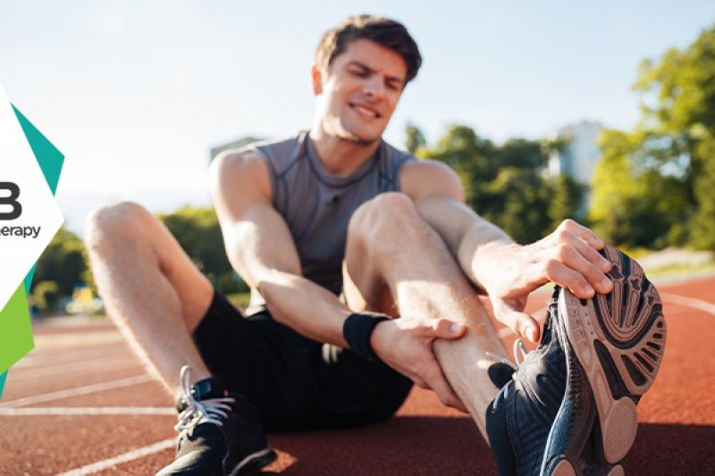
Rest: Avoid activities that stress the calf muscle. Crutches may be used to prevent putting weight on the injured leg.

Cryotherapy: Cryotherapy (ice packs) is applied to the affected area for 15-20 minutes every 1-2 hours to reduce swelling and numb the pain, wrapped in a cloth or towel as a barrier.

Compression: An elastic bandage or compression sleeve is used to minimize swelling. It should be ensured that it is not so tight that it restricts circulation.

Elevation: The injured leg is elevated above heart level when possible to reduce swelling.

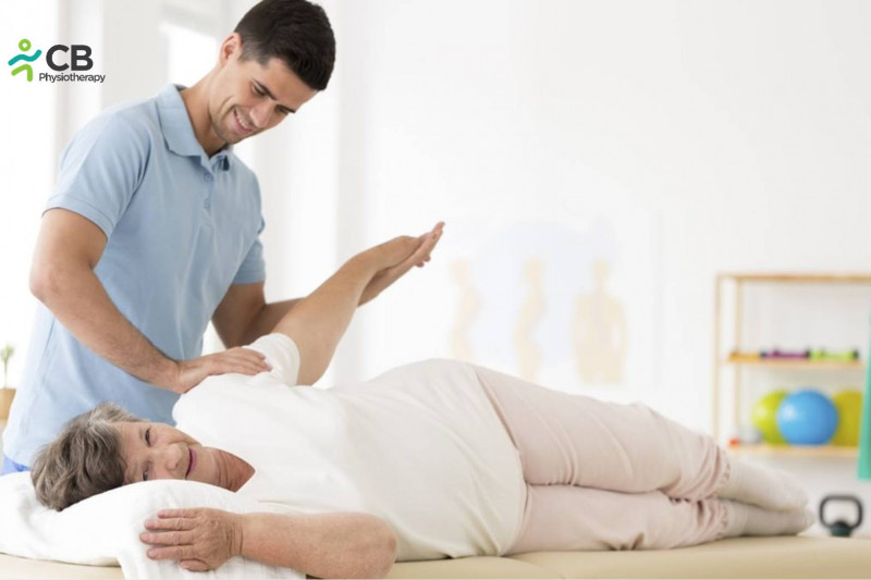
Exercises:
1. Gentle range-of-motion exercises are recommended to maintain mobility without stressing the muscle.
2. Stretching exercises focus on the calf muscle to prevent stiffness.

Balance and proprioception Training:
Help to improve stability and prevent re-injury like Sport-specific drills and activities help to restore full function.

Plyometric Exercises:
Plyometric exercises include jumping, sprinting, etc as tolerated to prepare for return to normal activity.

Massage Therapy:
Once the acute phase has passed, massage can help reduce muscle tightness and improve blood flow to the injured area.

Transcutaneous Electrical Nerve Stimulation (TENS):
1: Purpose: TENS is primarily used for pain relief. It works by sending low-voltage electrical currents through the skin to stimulate nerves and block pain signals from reaching the brain.
2: How It Works: Electrodes are placed on the skin near the site of the injury, and electrical impulses are delivered, which can help reduce pain by stimulating the release of endorphins (the body’s natural painkillers).
3: Benefits: Provides non-invasive pain management and can be used as often as needed.

Electrical Muscle Stimulation (EMS):
1: Purpose: EMS is used to stimulate muscle contractions, which can help prevent muscle atrophy, improve muscle strength, and promote blood circulation in the injured area.
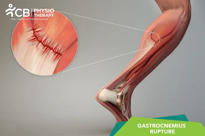
2: How It Works: Electrodes are placed on the skin over the gastrocnemius muscle, and electrical impulses are sent to cause muscle contractions. This can help maintain muscle tone during the recovery process.
3: Benefits: Enhances muscle recovery, reduces swelling, and can be particularly useful in the early stages of rehabilitation when active movement is limited.

Interferential Therapy (IFT):
1: Purpose: IFT is used for pain relief, reducing inflammation, and promoting healing. It involves the use of two high-frequency electrical currents that intersect within the tissue, creating a low-frequency current that penetrates deeper than TENS.
2: How It Works: Electrodes are placed around the injured area, and the intersecting currents create a therapeutic effect that helps reduce pain and swelling while promoting tissue healing.
3: Benefits: Effective for deeper tissue penetration and can be used to treat both acute and chronic pain associated with muscle tears.
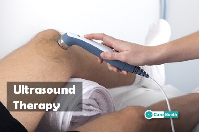
Ultrasound Therapy:
1: Purpose: Although not an electrical modality in the traditional sense, therapeutic ultrasound is often used in conjunction with other electrical therapies. It uses high-frequency sound waves to promote tissue healing, reduce inflammation, and break down scar tissue.
2: How It Works: A gel is applied to the skin over the gastrocnemius muscle, and a handheld ultrasound device is moved over the area. The sound waves penetrate the tissue, generating heat and promoting increased blood flow and tissue regeneration.
3: Benefits: Enhances healing of the muscle tissue and is particularly effective in the subacute phase of injury.

Low-Level Laser Therapy (LLLT):
1: Purpose: LLLT, also known as cold laser therapy, uses low-intensity light to promote healing at the cellular level, reduce pain, and decrease inflammation.
2: How It Works: The laser device emits light at specific wavelengths, which penetrates the skin and stimulates cellular activity, enhancing tissue repair and reducing inflammation.
3: Benefits: Non-invasive and effective in accelerating the healing process of soft tissue injuries like a gastrocnemius tear.

Neuromuscular Electrical Stimulation (NMES):
1: Purpose: Similar to EMS, NMES is used to stimulate muscle contractions but is more targeted at improving neuromuscular function and coordination, which can be beneficial during the later stages of rehabilitation.
2: How It Works: Electrodes are placed on the skin over the muscle, and electrical impulses are used to create controlled contractions, helping to retrain the muscle and improve its strength and endurance.
3: Benefits: Helps in muscle re-education, particularly important when returning to activities that require precise muscle control.

Pulsed Electromagnetic Field Therapy (PEMF):
1: Purpose: PEMF is used to enhance cellular repair and regeneration by applying electromagnetic fields to the affected area.
2: How It Works: The therapy involves placing a device that emits electromagnetic fields over the injured muscle, which penetrates the tissue and stimulates cellular repair processes.
3: Benefits: Non-invasive and helpful in reducing pain and inflammation while accelerating the healing process.

PATIENT EDUCATION.

Educating the patient about their gastrocnemius tear, including the nature of the injury, the importance of proper rehabilitation, and strategies for preventing future injuries, is essential for a successful recovery. Patient is educated to always warm up before physical activity to prepare the muscles for exercise. Regularly strengthen the calf muscles and maintain flexibility to reduce the risk of future tears. And is also advised to avoid sudden increases in the intensity or duration of physical activities.

Gastrocnemius Rupture Physiotherapy Nearby

Select your City to find & connect with our experts regarding Physiotherapy for Gastrocnemius Rupture

Related Conditions

conditions