smith-fracture.webp

Smith Fracture

Questions

WHAT IS OF SMITH FRACTURE?

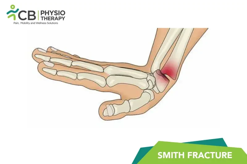
A Smith fracture, also known as a reverse Colles' fracture, is a fracture of the distal radius with volar (palmar) displacement of the distal fragment. It typically results from a fall onto a flexed wrist or a direct blow to the back of the wrist, causing the broken fragment of the radius to be displaced toward the palm side of the wrist.

WHAT ARE THE SIGNS AND SYMPTOMS OF SMITH FRACTURE?

The signs and symptoms of Smith fracture vary from patient to patient:

1: Pain: Immediate and severe pain in the wrist and forearm.
2: Swelling: Swelling around the wrist, which can extend to the hand and forearm.
3: Deformity: Visible deformity or an abnormal angle of the wrist, with the hand appearing displaced towards the pa
4: Bruising: Bruising or discoloration around the wrist.
5: Tenderness: Tenderness when touching the wrist or forearm.
6: Reduced Range of Motion: Difficulty or inability to move the wrist and fingers.
7: Numbness or Tingling: Numbness or tingling in the fingers if there is nerve involvement.

WHAT ARE THE CAUSES OF SMITH FRACTURE?

The causes of Smith fracture vary from:
1: Falls: Falling onto a flexed wrist is the most common cause, often occurring during sports or accidental slips and falls.
2: Direct Trauma: Direct impact to the back of the wrist, such as being hit by an object or in a car accident.
3: Osteoporosis: Weakened bones due to osteoporosis can be more susceptible to fractures, even with minor trauma.
4: Sports Injuries: Activities that involve a high risk of falls or direct wrist impacts, such as skiing, skateboarding, or contact sports.

 
Pathology
A Smith fracture involves a break in the distal radius bone with the distal fragment displaced towards the palm (volar displacement).

DIAGNOSIS OF SMITH FRACTURE.

Clinical Examination:
Physical examination to assess pain, swelling, deformity, and range of motion.
Neurological examination to check for nerve damage.

X-rays:
X-ray is the diagnostic tool, used to confirm the fracture, and determine the type and displacement of the fracture.

CT scan:
CT scan is used in complex fractures to get detailed images of bone fragments and joint involvement.

MRI:
MRI is done if there is suspicion of soft tissue, ligament, or nerve damage.

TREATMENT FOR SMITH FRACTURE.

Medication: Nonsteroidal Anti-Inflammatory Drugs (NSAIDs), Acetaminophen, Opioids, Antibiotics, etc.
(Note: Medication should not be taken without the doctor’s prescription.)

Surgical:
Surgical treatment for a Smith fracture, also known as a volar displaced distal radius fracture, is typically considered when non-surgical methods (like casting or splinting) are insufficient to properly align and stabilize the fracture. Here are the primary surgical techniques used to treat a Smith fracture:

1: Open Reduction and Internal Fixation (ORIF): Surgical procedure to realign the bones and secure them with plates, screws, or pins.

2: External Fixation: Used in cases with severe swelling or when internal fixation is not feasible, involving the use of an external frame to stabilize the bones.

PHYSIOTHERAPY TREATMENT FOR SMITH FRACTURE.

Immobilization:
The wrist is immobilized in a cast or splint for a period to ensure stability and promote healing.

Transcutaneous Electrical Nerve Stimulation (TENS)
Purpose: Pain relief.
Mechanism: TENS uses low-voltage electrical currents to stimulate nerves and block pain signals to the brain. It may also promote the release of endorphins, which are natural painkillers.
Application: Electrodes are placed on the skin around the fracture site. TENS can be used during or after immobilization and during rehabilitation.

Neuromuscular Electrical Stimulation (NMES)
Purpose: Prevent muscle atrophy and improve muscle strength.
Mechanism: NMES sends electrical impulses to stimulate muscle contractions, which helps maintain muscle mass and strength during periods of immobilization.
Application: Electrodes are placed on the skin over the muscles surrounding the fracture. NMES can be used after the initial healing phase when it is safe to begin muscle activation.

Interferential Current (IFC)
Purpose: Pain relief and reduction of swelling.
Mechanism: IFC uses two high-frequency currents that intersect to create a low-frequency stimulation deep within the tissues.
Application: Electrodes are placed around the fracture site. IFC is useful during both the acute and subacute phases of healing.

Wrist Mobilization:
Initiated after the initial healing phase to restore wrist function and regain the range of motion. Therapy usually starts with gentle movements and progresses to more intensive exercises as healing allows.
Gentle passive and active range of motion exercises for the wrist and forearm. This may include flexion, extension, supination, and pronation exercises.

Finger Exercises:
Gentle movements of the fingers to prevent stiffness and maintain circulation.

Elbow and Shoulder Exercises:
Range of motion exercises for the elbow and shoulder to prevent stiffness and maintain mobility.

Strengthening Exercises:
Gradual introduction of strengthening exercises for the wrist and forearm using resistance bands, light weights, or putty.

Advanced Strengthening:
Progress to more challenging strengthening exercises, including weight-bearing exercises and the use of heavier resistance.

Functional Activities:
Activities that mimic daily tasks to help restore functional use of the hand and wrist.
PATIENT EDUCATION.

Educating patients about their condition, treatment options, and recovery process is crucial for effective management and optimal recovery from a Smith fracture.

Smith Fracture Physiotherapy Nearby

Select your City to find & connect with our experts regarding Physiotherapy for Smith Fracture

Related Conditions