klumpke-s-palsy.webp

Klumpke's Palsy

Questions

WHAT IS KLUMPKE'S PALSY?

Klumpke's palsy is a form of brachial plexus injury that primarily affects the lower roots of the brachial plexus (C8 and T1). This condition leads to weakness or paralysis of the muscles in the forearm and hand.

WHAT ARE THE SIGNS AND SYMPTOMS OF KLUMPKE’S PALSY?

The signs and symptoms of Klumpke's palsy may vary from patient to patient, these may include:

1: Muscle Weakness or Paralysis: Particularly in the forearm and hand muscles.
2: Claw Hand Deformity: Due to paralysis of the intrinsic muscles of the hand, leading to hyperextension at the metacarpophalangeal joints and flexion at the interphalangeal joints.
3: Loss of Sensation: In the medial aspect of the forearm and hand.
4: Horner's Syndrome (in some cases): This may include ptosis (drooping eyelid), miosis (constricted pupil), and anhidrosis (loss of sweating) on the affected side of the face if the sympathetic fibers are involved.

WHAT ARE THE CAUSES OF KLUMPKE'S PALSY?

Klumpke's palsy can be caused by many reasons, a few of them are:

1: Birth Trauma: Excessive pulling on the infant's shoulder during delivery, especially in breech presentations.
2: Trauma: Direct injury or severe traction to the arm, such as in car accidents or falls.
3: Tumors: Compression from tumors in the neck or chest.
4: Inflammatory Conditions: Such as brachial plexitis.


Pathology

1: Nerve Root Avulsion or Stretching: Damage to the lower brachial plexus nerves (C8 and T1), which may be avulsed (pulled out from the spinal cord), stretched, or otherwise damaged.
2: Disruption of Nerve Function: This leads to loss of motor and sensory function in the muscles and skin innervated by these nerves.
3: Muscle Atrophy: Over time, the muscles innervated by the affected nerves may atrophy due to lack of stimulation.
4: Scarring and Fibrosis: Chronic injury may lead to scar tissue formation, further impeding nerve function and regeneration.

DIAGNOSIS OF KLUMPKE’S PALSY.

Diagnosing Klumpke's palsy involves a combination of clinical examination, imaging studies, and electrophysiological tests. Here are the primary techniques used:

Physical Examination:
Assessing muscle strength, sensation, and reflexes in the affected arm and hand. Looking for characteristic signs such as claw hand deformity and any signs of Horner's syndrome.

Magnetic Resonance Imaging (MRI):
MRI provides detailed images of the brachial plexus to detect nerve root avulsions, tumors, or other structural abnormalities.

Computed Tomography (CT) Myelography:
CT is useful in cases where MRI is inconclusive, especially for identifying nerve root avulsions.

Ultrasound:
Ultrasound can be used to visualize the brachial plexus, though it is less detailed than MRI.

Electromyography (EMG):
EMG measures the electrical activity of muscles and can help determine the extent and location of nerve damage.

Nerve Conduction Studies (NCS):
NCV helps to assess the speed and strength of electrical signals traveling through the nerves, helping to pinpoint the location and extent of nerve damage.

X-Rays:
X-rays are useful for detecting associated fractures or dislocations that might contribute to nerve damage.

Blood Tests:
Blood test helps to rule out any underlying conditions that might contribute to nerve damage, such as diabetes or inflammatory diseases.

TREATMENT FOR KLUMPKE'S PALSY.

Medication: Nonsteroidal Anti-Inflammatory Drugs (NSAIDs, Analgesics, Opioids, Anticonvulsants, Corticosteroids, Muscle Relaxants, etc.
(Note: Medication should not be taken without the doctor's prescription.)

Surgery:
Surgical intervention may be considered if there is no significant improvement with conservative treatments like physiotherapy. Surgery for Klumpke's palsy, which affects the lower nerves of the brachial plexus, aims to restore function to the forearm and hand. Here are the key surgical options:

Nerve Grafting:
It is a process in which grafting of healthy nerves from another body part is done to bridge damaged nerve gaps, it helps restore nerve function.

Nerve Transfers:
Nerve transfer is done for rerouting a healthy, less critical nerve to the damaged nerve, it helps improve function and strength.

Neurolysis:
Neurolysis is a process of removing scar tissue and compressing the nerve, it helps relieve pain and improves function.

Tendon Transfers:
Moving tendons from functioning muscles to paralyzed ones helps enhance hand and arm function.

Muscle Transfers:
Transferring muscles with their nerve supply from another body part helps improve muscle strength and functionality.

PHYSIOTHERAPY TREATMENT FOR KLUMPKE’S PARALYSIS.

Thermotherapy/ cryotherapy:
Heat and cold therapy help manage pain and inflammation.

Transcutaneous Electrical Nerve Stimulation (TENS):
Tens is used for pain relief. It delivers low-voltage electrical currents through the skin to stimulate nerves and reduce pain signals to the brain.

Neuromuscular Electrical Stimulation (NMES):
a) NMES is used to stimulate muscle contractions and improve muscle strength and function.
b) It delivers electrical impulses to motor nerves, causing muscles to contract. The electrodes are placed on the skin over the targeted muscles to facilitate contractions and promote muscle re-education.

Functional Electrical Stimulation (FES):
a) FES helps enhance functional movement by stimulating muscles during specific activities.
b) Mechanism: It delivers electrical impulses in a coordinated pattern to support voluntary movement and functional tasks. It is often used during activities like walking, grasping, or reaching to improve functional use of the affected limb.

Interferential Current Therapy (IFC):
IFT helps in pain relief and reduction of inflammation. It uses medium-frequency electrical currents that intersect below the skin to provide deeper tissue stimulation. The electrodes are positioned around the area of pain, with the currents intersecting at the target tissue.
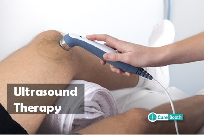
Microcurrent Therapy:
Microcurrent therapy helps promote healing and reduce pain. It uses very low-level electrical currents to stimulate cellular repair and reduce inflammation. The electrodes are placed on the skin over the affected area.

Iontophoresis:
Iontophoresis helps deliver medication through the skin using electrical currents. It uses a small electric current to drive charged medication molecules through the skin to the underlying tissue. It helps for localized drug delivery, often to reduce inflammation or pain.

Galvanic Stimulation:
Galvanic stimulation helps promote blood flow and healing. It uses direct current to stimulate tissues and increase blood flow to the area. The electrodes are placed on the skin over the affected area.

Range of Motion Exercises:
a) Passive Range of Motion (PROM):
These exercises are performed with the help of the therapist to maintain joint flexibility and prevent stiffness when the patient cannot move the limb actively.
b) Active-Assisted Range of Motion (AAROM):
The patient attempts to move the affected limb with the help of the therapist or using the unaffected limb.
c) Active Range of Motion (AROM):
The patient moves the limb independently to improve joint mobility and muscle strength.

Strengthening Exercises:
a) Isometric Exercises:
These involve contracting the muscles without moving the joint, which helps to maintain muscle strength without putting strain on the injured nerves.
b) Resistance Training:
Gradually incorporate resistance bands or light weights to strengthen the muscles of the forearm and hand as nerve function improves.

Stretching Exercises:
a) Gentle Stretching:
To maintain flexibility in the muscles and tendons and to prevent contractures.
b) Specific Stretches:
Targeted at the muscles of the forearm and hand to maintain a full range of motion in the joints.

Functional Training:
a) Task-Oriented Exercises:
Focused on improving the ability to perform daily activities, such as grasping, holding, and manipulating objects.
b) Fine Motor Skills:
Exercises to improve dexterity and coordination in the fingers and hands.

Sensory Re-education:
a) Desensitization Techniques:
Using different textures and temperatures to help the nerves adapt to sensory input and reduce hypersensitivity.
b) Tactile Stimulation:
Activities that stimulate sensory feedback to improve sensation in the affected areas.
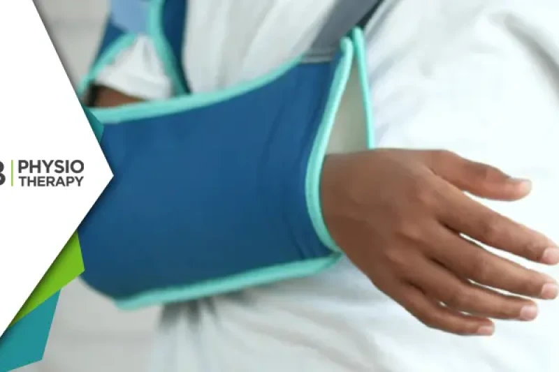
Splinting and Orthotics:
Splints: To support the wrist and hand in a functional position, prevent deformities, and improve alignment.
Dynamic Splints: Allowing for some movement while providing support to weak muscles.
Manual Therapy: Including gentle massage and mobilization techniques to reduce pain and improve circulation.

PATIENT EDUCATION

Patient education involves teaching proper techniques for exercises, safe ways to perform daily activities, and the importance of adherence to the therapy plan. The patient is advised to perform exercises at home to ensure continuity of care and progress. Recommendations are also given for modifications at home or work to facilitate ease of movement and reduce strain.

Klumpke's Palsy Physiotherapy Nearby

Select your City to find & connect with our experts regarding Physiotherapy for Klumpke's Palsy