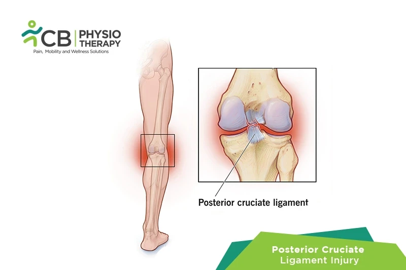
The posterior cruciate ligament (PCL) and the anterior cruciate ligament (ACL) connect the thighbone or femur to the shinbone i.e tibia. The anterior and posterior cruciate ligaments both form an "X" in the center of the knee. If one of these ligaments is torn, then it might cause pain, swelling, and a feeling of instability. Though posterior cruciate ligament (PCL) injury occurs far less often than the anterior cruciate ligament (ACL) if in case it is injured, it causes less pain, disability, and knee instability than an ACL tear, but still takes several weeks or months to recover.
PCL injuries are graded according to severity:
Grade 1: There is limited damage as a result of overstretching. Knee stability and function are not affected.
Grade 2: In this grade, the ligament is partially torn and the patient feels instability.
Grade 3: The patient has a complete ligament tear or rupture. This type of injury is usually accompanied by a sprain of the ACL or/ and collateral ligaments.
If only the posterior cruciate ligament is injured then the symptoms will be so mild they are not noticed, but after some time, the pain might worsen and the knee might feel more unstable. But if other parts of the knee have also been injured, then signs and symptoms are more severe. The patient feels:
The posterior cruciate ligament can be torn alone, or along with the other ligament. This can be due to:
Physical examination:
Physical examination is done by palpating, the looseness or fluid in the joint. The patient is asked to move the knee, leg, or foot in different directions also asked to stand and walk. The injured leg is compared with the healthy leg to look for sagging or abnormal movement in the knee or shinbone.
X-ray:
An X-ray cannot detect ligament damage but can show bone fractures. Posterior cruciate ligament injuries sometimes have breaks in which a small chunk of bone, is pulled away from the main bone. It can be done in different positions, like standing and weight-bearing with 45° knee flexion.
Magnetic resonance imaging (MRI) scan:
Magnetic resonance imaging (MRI) scan is a painless procedure that uses radio waves and a strong magnetic field to produce images of the soft tissues of the body. MRI scan can show a posterior cruciate ligament tear and also determines whether the other knee ligaments or cartilage are injured. It may appear normal in grade I and II injures.
Medications: Anti-inflammatory medications, analgesics, pain killers, etc.
Note: Medication should not be taken without the doctor's prescription.
Surgery
Treatment depends on the posterior cruciate ligament (PCL) injury on the extent of the injury, in most cases, surgery isn't required.
In severe cases, if the injury is combined with other torn knee ligaments, cartilage damage, or a broken bone, then surgery to reconstruct the ligament might be required. Surgery might also be considered if there are continuous episodes of knee instability despite appropriate rehabilitation.
Arthroscopy: It is a surgical technique, to look inside the knee joint. A tiny video camera is inserted into the knee joint through a small incision, to view the images of the inside of the joint on a computer monitor or TV screen.
Reconstruction: Posterior Cruciate Ligament reconstruction is done to restore normal knee mechanics and dynamic knee stability, and thus correct posterior tibial laxity.
Rest:
Stay off the injured knee, to protect it from further damage. For mobility, crutches are used.
Ice packs are applied to the knee for 20 to 30 minutes after every 3 to 4 hours for 2 to 3 days.
Compression:
For compression, an elastic bandage is wrapped around the knee.
Elevation:
To elevate lie down and place a pillow under the knee to help reduce swelling.
Ultrasound therapy is used to reduce pain and increase the range of motion.
Transcutaneous electrical stimulation (TENS):
Transcutaneous electrical stimulations are used to decrease pain and swelling.
Range of motion exercises is used to help decrease swelling and restore the range of motion of the knee.
Stretching exercises:
Stretching exercises are held for 30-seconds, followed by relaxation and repeated 5 times, these stretches can be given to hamstrings, iliotibial tract, gastrocsoleus, etc.
Strengthening exercises include isometrics, isotonic and isokinetic exercises. Quad sets, hams sets, hamstring curls, straight leg raises, heel raises, heel dig bridging, shallow standing knee bends are examples of strengthening exercises.
Progressive resistive exercises (PRE):
Progressive resistive exercises are self-resistive exercises that are progressed gradually to self-generated tension and resistance exercise with weight belts.
Balance exercises:
Balance exercises are used to enhance balance and equilibrium by using a bobath ball or a wobble board.
Plyometrics:
Plyometric exercises like high-intensity exercises are used to meet the high demands of strenuous and sports activities. These high-intensity exercises include squat jumps, clapping pushups, reverse lunge knee-ups, jumping, running, etc.
The patient is asked to avoid twisting or pivoting movement of the knee. Strengthening and stretching exercises should be continued to prevent reinjury. The patient should be careful while doing strenuous activities.
Select your City to find & connect with our experts regarding Physiotherapy for Posterior Cruciate Ligament(pcl) Injury