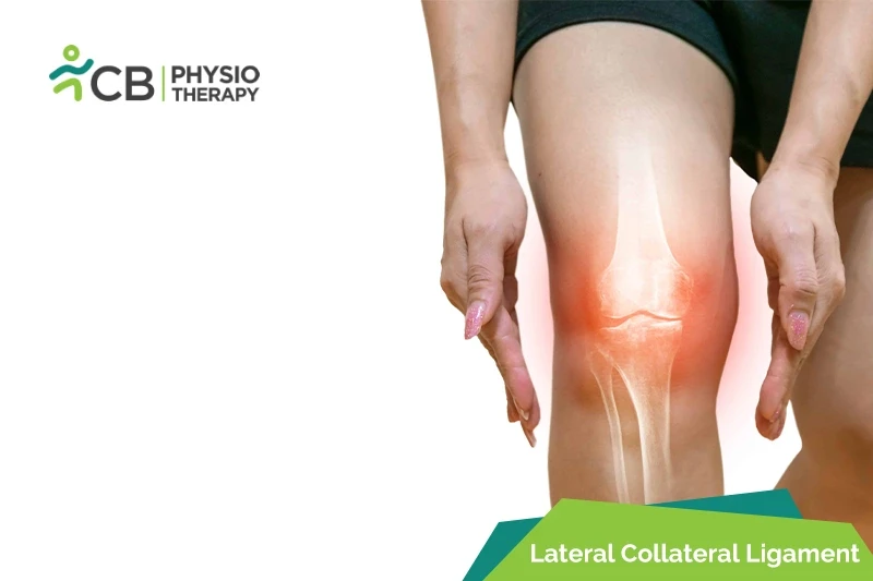
घुटने का जोड़ शरीर का सबसे बड़ा जोड़ है और चोटिल होने के लिए सबसे आसान जोड़ों में से एक है। पार्श्व संपार्श्विक बंधन (एलसीएल) मोच और आंसू, हालांकि, घुटने की सामान्य चोटें बहुत कम हैं। घुटने के संपार्श्विक स्नायुबंधन पार्श्व संपार्श्विक बंधन (LCL) और औसत दर्जे का संपार्श्विक बंधन (MCL) हैं, जो घुटने के दोनों ओर स्थित होते हैं। ये स्नायुबंधन संयुक्त की बग़ल में गति को नियंत्रित करते हैं, इसे अगल-बगल की असामान्य गति से बचाते हैं। पार्श्व संपार्श्विक बंधन (एलसीएल) घुटने के बाहरी हिस्से के साथ चलता है।
पार्श्व संपार्श्विक बंधन (LCL) मोच के प्रकार:
ग्रेड 1 लेटरल कोलेटरल लिगामेंट (LCL) मोच, लिगामेंट फैला हुआ है, हल्के से मध्यम दर्द के साथ, हल्की चोट , और गति की न्यूनतम कमी हुई सीमा। लेकिन अभी भी घुटने को स्थिर और क्रियाशील रखने में सक्षम है।
ग्रेड 2 लेटरल कोलेटरल लिगामेंट (LCL) मोच या आंशिक LCL फटना, गंभीर दर्द के साथ, लिगामेंट में खिंचाव या तनावग्रस्त हो जाता है और ढीला हो जाता है और गति की सीमा मध्यम से गंभीर कम हो जाती है।
ग्रेड 3 लेटरल कोलैटरल लिगामेंट (LCL) मोच या गंभीर दर्द के साथ पूरा LCL टूटना, लिगामेंट दो हिस्सों में फट जाता है अलग-अलग टुकड़े और जोड़ अब स्थिर नहीं है और गति की सीमा गंभीर रूप से कम हो गई है।
घुटने का जोड़ कठोर मांसपेशियों के संकुचन और प्रत्यक्ष प्रभाव के प्रति संवेदनशील होता है, जिसके कारण होता है:
<उल शैली = "सूची-शैली-प्रकार: डिस्क;">
लेटरल कोलेटरल लिगामेंट (LCL) मोच के लक्षण गंभीरता में भिन्न हो सकते हैं, जो चोट की श्रेणी पर निर्भर करता है। लक्षणों में शामिल हैं:
<उल शैली = "सूची-शैली-प्रकार: डिस्क;">
पैथोलॉजी:
लेटरल कोलैटरल लिगामेंट इंजरी उस कनेक्शन का व्यवधान है जो लेटरल फीमर और लेटरल के बीच मौजूद है बहिर्जंघिका। एक पार्श्व संपार्श्विक मोच को I, II और III डिग्री के रूप में वर्गीकृत किया गया है। यह कुछ या बिना किसी ढिलाई के, निरंतरता में स्नायुबंधन के साथ, या पूर्ण व्यवधान के साथ हल्की चोट का कारण बन सकता है।
शारीरिक परीक्षा:
शारीरिक परीक्षा में पार्श्व संयुक्त रेखा के साथ सूजन, इकोस्मोसिस और गर्मी का निरीक्षण करना शामिल है। रोगी को पार्श्व संयुक्त रेखा के साथ गति, मांसपेशियों की ताकत, सनसनी, सजगता और तालु की सीमा के लिए मूल्यांकन किया जाता है। संबंधित लिगामेंटस, मेनिस्कल, या सॉफ्ट टिश्यू इंजरी को निर्धारित करने के लिए गैट विश्लेषण और विशेष परीक्षण किए जाते हैं।
एक्स-रे:
चुंबकीय अनुनाद इमेजिंग (MRI):
चुंबकीय अनुनाद इमेजिंग (MRI) LCL चोटों के निदान में स्वर्ण मानक है। LCL चोट के निदान में कोरोनल और सजिटल छवियों का उपयोग किया जाता है।
LCL चोट के त्वरित निदान के मामले में अल्ट्रासाउंड का उपयोग किया जाता है। गाढ़ा और हाइपोचोइक एलसीएल एलसीएल की चोट का संकेत देता है। पूर्ण रूप से फटने की स्थिति में, अल्ट्रासाउंड में एडिमा में वृद्धि, फाइबर निरंतरता की कमी, LCL की गतिशील शिथिलता दिखाई दे सकती है।
ध्यान दें: डॉक्टर के नुस्खे के बिना दवा नहीं लेनी चाहिए।
सर्जरी:
लेटरल कोलेटरल लिगामेंट (LCL) चोटें घुटने की अन्य संरचनाओं को शामिल करती हैं जिन्हें अतिरिक्त उपचार की आवश्यकता हो सकती है। कुछ एलसीएल मोच रूढ़िवादी उपचार से ठीक हो सकते हैं दूसरों को सर्जरी की आवश्यकता होती है।
सर्जरी आर्थ्रोस्कोपी के साथ एक खुली प्रक्रिया के रूप में की जाती है। फटे हुए LCL को एक टिश्यू ग्राफ्ट का उपयोग करके बदल दिया जाता है, जिसे हड्डी के भीतर सुरंगों के माध्यम से पारित किया जाता है और फिर स्क्रू का उपयोग करके फाइबुला और फीमर हड्डियों से जोड़ा जाता है।
पुनर्निर्माण एक अन्य तकनीक है जिसका उपयोग किया जाता है, इसमें LCL के पुनर्निर्माण के लिए एलोग्राफ़्ट हैमस्ट्रिंग टेंडन या एक ऑटोग्राफ शामिल होता है। आमतौर पर, पटेलर टेंडन एलोग्राफ़्ट के साथ घुटने में LCL का पुनर्निर्माण भी किया जाता है।
आराम:
घायल क्षेत्र को किसी भी तनाव से बचाने और इसे आगे की चोट से बचाने के लिए आराम की सलाह दी जाती है। यह नी ब्रेस, लिगामेंट को आगे साइडवे मूवमेंट से बचाने में मददगार हो सकता है और उपचार प्रक्रिया को गति देने में भी मदद कर सकता है।
घायल क्षेत्र पर चोट लगने के तुरंत बाद हर घंटे में 15 से 20 मिनट के लिए बर्फ लगाने से जोड़ों की सूजन और सूजन को कम करने में मदद मिल सकती है।
संपीड़न:
क्रायोथेरेपी के बाद, एक कम्प्रेशन स्लीव पहना जाता है, यह सूजन को नियंत्रित करने और घुटने की सुरक्षा करने में मदद करता है।
ऊंचाई:
अल्ट्रासाउंड थेरेपी का उपयोग दर्द को कम करने और गति की सीमा को बढ़ाने के लिए किया जाता है।
ट्रांसक्यूटेनियस इलेक्ट्रिकल स्टिम्युलेशन (TENS):
ट्रांसक्यूटेनियस इलेक्ट्रिकल स्टिमुलेशन (TENS) का उपयोग दर्द और सूजन को कम करने के लिए किया जाता है।
गति अभ्यास की सीमा:
फिजियोथेरेपी घुटने के आसपास की मांसपेशियों को मजबूत करने में मदद करती है। उदा. एक प्रतिरोध बैंड लें और इसे निचले पैर पर रखें, पैरों को कूल्हे-चौड़ाई से अलग रखा जाना चाहिए, फिर नीचे झुकें और एक पैर को कूल्हे-चौड़ाई से चौड़ा करके बाहर निकलें, फिर दूसरे पैर को पीछे लाएं, कूल्हे-चौड़ाई पर लौटें। इस अभ्यास को प्रत्येक दिशा में 10 बार दोहराएं और दर्द रहित गति की सीमा में किया जाना चाहिए।
स्ट्रेचिंग एक्सरसाइज:
प्रगतिशील प्रतिरोधक व्यायाम (PRE):
संतुलन अभ्यास:
संतुलन अभ्यास एक डगमगाने वाले बोर्ड की मदद से किया जाता है जिसका उपयोग संतुलन और संतुलन को बढ़ाने के लिए किया जाता है।
प्लायोमेट्रिक्स:
प्लायोमेट्रिक अभ्यास उच्च-तीव्रता वाले व्यायाम हैं जिनका उपयोग खेल गतिविधियों जैसी उच्च माँगों से निपटने के लिए किया जाता है। उदाहरण हैं कूदना, दौड़ना, रिवर्स लंज नी-अप्स, स्क्वाट जंप्स, क्लैपिंग पुश-अप्स आदि।
रोगी को सलाह दी जाती है कि घुटने को फिर से होने से रोकने के लिए मज़बूत व्यायाम करना जारी रखें -चोट। गतिविधियों के दौरान रोगी ब्रेस पहनना जारी रख सकता है।
फिजियोथेरेपी के बारे में हमारे विशेषज्ञों को खोजने और उनसे जुड़ने के लिए अपने शहर का चयन करें पार्श्व संपार्श्विक बंधन (एलसीएल) चोट