hemangioma.webp

Hemangioma

Questions

WHAT IS HEMANGIOMA?

Hemangioma is a common benign (non-cancerous) growth of blood vessels. It typically appears as a red or purplish birthmark on the skin. Hemangiomas can occur anywhere on the body but are most often found on the face, head, neck, spine, or any other part of the body. They can vary in size, shape, and color.

WHAT ARE THE SYMPTOMS OF HEMANGIOMA?

The symptoms of hemangioma vary from person to person, depending on the severity:

1: Appearance: Hemangiomas usually start as small, flat, red marks on the skin. They can be mistaken for a simple red birthmark or rash in their early stages. However, they often grow in size and may change in appearance over time.
2: Growth: Hemangiomas are characterized by rapid growth during the first few months of life. They may continue to grow for up to a year before eventually stabilizing.
3: Color Changes: They can change in color, typically from bright red to purplish blue, before eventually fading away.
4: Texture: The texture of the skin over a hemangioma might be different from the surrounding skin. It can be raised, bumpy, or have a spongy texture.
5: Location: Hemangiomas can occur anywhere on the body but are most commonly found on the face, head, or neck.
6: Complications: In some cases, hemangiomas can cause complications, such as ulceration, bleeding, infection, or interference with vision or breathing if located in certain areas.

WHAT ARE THE CAUSES OF HEMANGIOMA?

The exact cause of hemangiomas is not entirely understood, but they are believed to result from an overgrowth of blood vessels. Some contributing factors and characteristics include:
1: Genetics: There may be a genetic predisposition to developing hemangiomas, as they tend to run in families.
2: Hormones: Hemangiomas are more common in females and premature infants, suggesting a possible hormonal influence.
3: Developmental Aberrations: Hemangiomas often appear in the first few weeks or months of life, suggesting that they may be related to an abnormality in the development of blood vessels.
4: Immune System: Some researchers believe that the immune system may play a role in the development of hemangiomas.
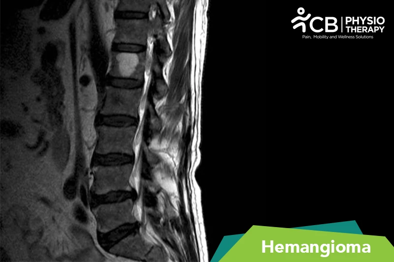

Pathology

The pathology of hemangioma involves the growth of abnormal blood vessels.
Hemangiomas are typically classified into two main types based on their appearance and behavior: infantile hemangiomas and congenital hemangiomas.

Infantile Hemangioma:
Infantile hemangiomas are the most common type and have a characteristic growth pattern.
They typically appear after birth and go through a growth phase followed by a regression phase. The growth phase involves the rapid proliferation of blood vessels, resulting in a raised, red, or purplish lesion on the skin. The regression phase involves the gradual shrinking and fading of the lesion. Histologically, infantile hemangiomas are composed of a disorganized mass of blood vessels, including capillaries and larger vessels. They are often characterized by endothelial cells, pericytes, and a fibrous component.

Congenital Hemangioma:
Congenital hemangiomas are present at birth and do not go through the same rapid growth and regression phases as infantile hemangiomas. They may be fully formed at birth or continue to grow at a slower rate. Congenital hemangiomas are often composed of mature blood vessels and do not have the same rapid proliferation as infantile hemangiomas.

DIAGNOSIS OF HEMANGIOMA.

Clinical Examination: Healthcare providers, including pediatricians and dermatologists, can often diagnose hemangiomas through a visual examination. The characteristic appearance of a hemangioma, with its red-to-purplishcolor and raised texture, is usually sufficient for diagnosis.

Dermoscopy: In some cases, a dermoscope, which is a handheld magnifying device, may be used to examine the lesion more closely and confirm the diagnosis.

Ultrasound: Ultrasound can provide information about the size and depth of the hemangioma and its relationship to surrounding structures.

MRI (Magnetic Resonance Imaging): MRI is more detailed than ultrasound and may be used when deeper tissue involvement is suspected or when there are concerns about potential complications.

Biopsy: In rare cases, a biopsy may be performed, especially if there is doubt about the diagnosis or if there is concern about a different condition. However, biopsies are generally avoided if possible because they can cause bleeding in the case of hemangiomas.

TREATMENT FOR HEMANGIOMA.

Medication: Beta-blockers (e.g., propranolol) or corticosteroids, etc.
Note: Medication should not be taken without the doctor’s prescription.

Pulsed Dye Laser Therapy: Laser therapy is a common treatment for hemangiomas, especially those on the skin's surface. Pulsed dye lasers work by targeting and coagulating the blood vessels in the hemangioma. This can help to shrink the lesion and reduce redness.

Surgical Excision: Surgical removal of a hemangioma may be considered in cases where it poses a significant risk, is disfiguring, or causes functional problems. This is typically reserved for older children or adults with residual hemangiomas that have not regressed.

Embolization: In some cases, particularly when hemangiomas are in internal organs or deep tissues, embolization may be necessary. This procedure involves blocking the blood vessels that supply the hemangioma.

Interferon Therapy: Interferon is an antiviral and immune-modulating medication that may be used for certain cases of hemangiomas that are resistant to other treatments. However, it is less commonly used due to potential side effects.

WHAT IS THE PHYSIOTHERAPY TREATMENT FOR HEMANGIOMA?

Thermotherapy: Thermotherapy involves using heat to treat hemangiomas which help relieve pain and increase circulation.

Transcutaneous electrical nerve stimulation (TENS): It works by applying electrical stimulation to the skin through electrodes placed on or near the area of pain, which may lead to the release of endorphins (natural pain relievers) and potentially block pain signals to the brain.

Lymphatic Drainage: If a hemangioma is in a location where it may impact lymphatic flow, physiotherapists may perform manual lymphatic drainage techniques to reduce swelling and improve lymphatic circulation. This can be particularly useful in cases of extensive hemangiomas.

Wound Care: In cases where hemangiomas have ulcerated or caused open wounds, physiotherapists can provide wound care and promote wound healing. This may involve wound cleaning, dressing changes, and instruction on wound care to prevent infection and complications.

Scar Management: Physiotherapists can provide scar management techniques to improve the flexibility, appearance, and texture of the skin in areas affected by hemangiomas. This may include scar massage, stretching exercises, and the use of silicone gel sheets or other scar management products.

Range of Motion Exercises: Hemangiomas, particularly those on or near joints, can lead to restricted joint movement. Physiotherapists can design exercise programs to improve joint range of motion and prevent contractures.

Strength and Conditioning: In cases where hemangiomas have affected the use of limbs or resulted in muscle weakness, physiotherapists can develop strength and conditioning programs to help patients regain muscle strength and functional abilities.

Orthotic or Assistive Devices: Physiotherapists can assess the need for and provide guidance on the use of orthotic devices, braces, or assistive equipment to enhance mobility and function for patients with hemangiomas affecting their musculoskeletal system.

Lymphedema Management: In some cases, particularly when hemangiomas are in areas that may impact lymphatic drainage, physiotherapists can provide lymphedema management techniques to reduce swelling and improve lymphatic circulation.

Functional Rehabilitation: For individuals with large or problematic hemangiomas that have led to physical disabilities or deformities, physiotherapists can provide rehabilitation programs to enhance functional abilities and improve overall well-being.

PATIENT EDUCATION.

The patient is educated about how to care for haemangiomas, including keeping the area clean and monitoring for signs of complications. The potential emotional impact of hemangiomas, especially in visible or problematic locations is discussed with the patient.The patient is educated about the importance of regular follow-up appointments with the physiotherapist to monitor the hemangioma's progress and adjust the treatment plan as necessary. Also, guidance is offered on protecting the hemangioma from injury, especially if it is in an area prone to trauma.

Hemangioma Physiotherapy Nearby

Select your City to find & connect with our experts regarding Physiotherapy for Hemangioma