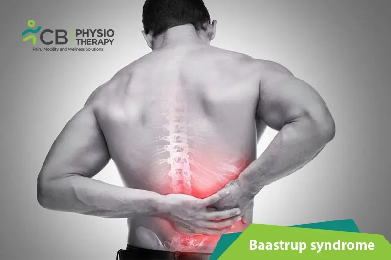
Baastrup's disease or Baastrup syndrome is a relatively common pathology affecting vertebral column. First formally described by Christian Ingerslev Baastrup in 1933, the disorder is characterized by the close approximation and contact of the spinous processes of adjacent vertebrae in the setting of degenerative spine disease. As a product of radio-graphic findings of this process, the syndrome is often referred to as “kissing disease”.
Baastrup Syndrome is more common amongst elderly persons, but this does not preclude incidence in younger individuals. The effect of gender is still unknown, so further research is necessary
· Excessive lordosis (excessive inward curvature of spine)
· Back pain more specifically, midline pain , that radiates distally and proximally, increasing on extension and reducing on flexion.
· Pain upon palpation at the level of pathological interspinous ligament.
· Inflammation
· Cystic lesions
· Sclerosis
· Flattening and enlargement of articulating surfaces
· Bursitis. ( inflammation of the sac like cavity, especially one countering friction at a joint)
Age is the most important factor responsible for the evolution of Baastrup syndrome. Other risk factors are:
· Excessive lordosis which results in increased mechanical pressure
· Repetitive strains of the interspinous ligament with subsequent degeneration and collapse
· Incorrect posture
· Traumatic injuries
· Tuberculous spondylitis
· Bilateral forms of congenital hip dislocation
· Stiffening of the thoracic spine or the thoracolumbar transition
· Obesity
The cause of pain is described as being mainly mechanical due to the neighbouring spinous processes coming into contact. Pain worsens with hyperextension or increased lordosis which can been seen in patients with obesity, limitation in hip movements and pro/elite swimmers.
Diagnosis: Diagnosis of Baastrup’s disease is verified with clinical examination as well as imaging studies.
Examination: Symptoms include low back pain with midline distribution that exacerbates when performing extension, relieved during flexion and is exaggerated upon finger pressure at the level of the pathologic interspinous ligament. Rotation and lateral flexion are also very painful. The pain can be described as a sharp or deep ache, often worse during physical activities that increase lumbar lordosis or compression of these structures.
Throughout the physical examination, the physiotherapist uses active and passive techniques with the intention of evoking complaints. Active spinal extension can reproduce the symptoms. The stork test is very beneficial in the examination of this disease. When the patient bends forward, relief is also gained.
Imaging modalities: Imaging modalities are required to avoid misdiagnosis. Some of the imaging modalities are:
· Computed tomography (CT) scan.
· Radiography (X-rays).
· Magnetic resonance imaging (MRI).
Medical management: Medical treatment can be conservative or surgical and an accurate diagnosis of the disease is necessary for determining appropriate treatment. Where an MRI shows active inflammatory changes or oedema, localised injections can be tried. If injections do not improve the patient's symptoms, surgical treatment is then recommended.
Non-surgical treatment consists of localised injections of analgesics or NSAIDS. which can be given bi-weekly. During this treatment period, extension movements of the lumbar spine should be avoided. After local anesthesia of the skin and subcutaneous tissues, the injection is given the painful interspinous ligaments between the affected spinous processes' under fluoroscopic control.
Suggested surgical therapies include: excision of the bursa, partial or total removal of the spinous process, or an osteotomy
Physical therapy management: The main goal is the reduction of pain as well as hyperlordosis and to improve spinal function. Once the pain is managed, physical therapy management can begin, involving education, strengthening and stretching of the abdominal and spinal muscles.
When the abdominal muscles are weak, the hip flexors are mainly responsible in shaping the lumbar spine. Furthermore the rectus femoris muscle is a continuation of the hip flexor complex so it is important to stretch these muscles.
Motion of the gluteus maximus muscle during the flexion-extension cycle is decreased in patients with chronic low back pain, which is why strengthening of this muscle should be part of the physical management program.
Physical therapy is also suggested to be helpful for reducing the neuromuscular damage that is provoked by the disease and other treatments such as, heat therapy.
Select your City to find & connect with our experts regarding Physiotherapy for Baastrup Syndrome